
Controlled damage
of
subcellular structures
We used optical scalpel to study a damage of living cells. These
experiments
were performed in cooperation with the group of Prof. R. Janisch from the Faculty of Medicine of the
Masaryk University in Brno. The effects of pulse
laser irradiation
were used
to study of the infusorians Paramecium
tetraurelia
and Blepharisma undulans
undulans.
Destruction of the
cytoskeleton by laser irradiation was detected by immunofluorescence
staining1. A difference in the development and
healing of
the wound
was observed between Paramecium
and Blepharisma cells.
A more
immediate reaction was
recorded in Blepharisma cells
containing blepharismin,
a red pigment, known to absorb light energy. The damage to the infusorian cortex due to laser
irradiation was
compared
with that produced by mechanical devices.
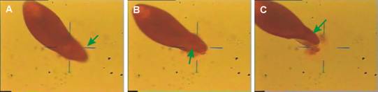
Fig. 1 Expresure
of the Blepharisma
cortex to a laser beam.
Place of hitting marks with green
pointer.
Experiments:
The sizes of
these protozoa
range from tens to hundreds of micrometers. Therefore they were immobilized by sucking into a microcapillary
(TransferMan NK
mechanical micromanipulator with a CellTram
Air micropipette
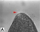
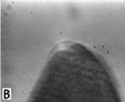
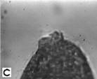
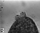
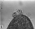
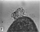
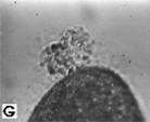
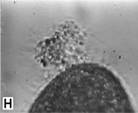
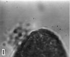
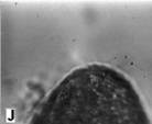
Fig. 2 Irradiation of the Blepharisma in detail. Place of hitting marks with red pointer.
Time span between A and J pictures was 8 sec.
Laser
Minilite II (Continuum)
was used to
generate UV laser pulse
with the following properties: wavelength,
l=355 nm; maximal pulse energy, 8 mJ;
pulse
length, 5 ns. The laser beam was enlarged by a system of lenses,
directed to an
Olympus IX70 microscope by dichroic
mirrors and
focused by Olympus Plan 20X and Plan 40X microscope objectives. The
position of
the beam in the viewing field of the microscope was adjusted laterally
and
longitudinally by movable lenses2,3.
Specific areas of the surface or selected structures close below the
surface of
an immobilized cell were irradiated by laser pulses of energy ranging
from 4 to
15 mJ. Influences of
the laser pulses on
selected cytoplasmic
structures were studied together with the reparation of damaged
cytoskeleton
using imunofluorescent
methods.
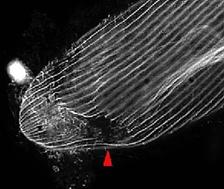
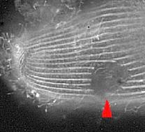
Fig. 3 Laser-induced wound in Blepharisma cortex visualized by immunofluorescence staining.
Defect in
cytosceleton
is marked by red arrow.
Results:
The response of Paramecium
tetraurelia to
the damage caused
by a laser is different comparing to the response to mechanical damage.
The
margins of the wound contract rapidly and reparation rate of membrane
decreases due
to local temperature increase. The cell of Blepharisma
undulans
undulans
contains a red
pigment
in the margin layer which absorbs laser energy better and therefore the cell
damage by
the pulse has more serious consequence comparing to the Paramecium
tetraurelia.
The increased
reproduction rate is an interesting result of cell damage.
References:
1. Z. Moravčík, R. Janisch, J. Ježek, P. Zemánek, “Response of infusorian cells to injury caused by a laser microbeam.” Scripta Medica 76, pp. 149-162, 2003. ABSTRACT
2. P. Zemánek, L. Šrámek,
A. Jonáš,
Z. Moravčík,
R. Janisch, M. Liška,
“Standing
wave trap and single beam gradient optical trap - experiments and
biological
applications.” Proc.
SPIE 3820, pp.
401–410, 1999. ABSTRACT
3. J. Ježek, A. Jonáš,
M. Liška, P.
Jedlička,
E. Lukášová,
S. Kozubek,
P. Zemanek. “Combined
system for optical cutting and multiple-beam optical
trapping.”
Proc SPIE
4016,
303–308, 2000.
ABSTRACT
Send comments to webmaster
Last modification: 30 Mar 2007