
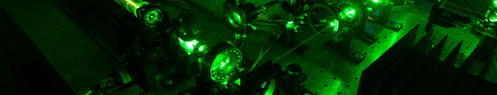


Microscopy and Spectroscopy of Surfaces
The group is engaged in scanning electron microscopy using slow and very slow electrons. Its activities are oriented toward the methodology, instrumental principles and compilation of unique elements of instrumentation for achieving a high resolution of images at electron energies down to units of electronvolts, and applications of low-energy microscopy in selected tasks in materials sciences and biomedical sciences. Its goal consists of the introduction and development of methods based on variability in the energy of the electron beam along the microscope column and the utilization of electron optical elements with a retarding field. The important issues it considers include detection of electrons emitted with very low energy, image resolution at low energies, procedures for sample preparation, vacuum issues, the acquisition of image data and the interpretation of micrographs. Surfaces of bulk samples are examined by means of the reflection of slow electrons, and ultrathin films studied by means of the passage of electrons. The main application areas include study of very clean and well defined surfaces, including surfaces treated in-situ in the ultrahigh vacuum space, observation of real surfaces under standard vacuum conditions, observation of nonconductive samples, and confrontation of image signals with electron-spectroscopic signals such as Auger electrons. Slow electrons enable us to image the grains in polycrystalline materials at high contrast, including the distribution of internal strain, thin surface layers at high sensitivity in respect of their morphology, doped areas in semiconductors at a high contrast proportional to the dopant density, ultrathin tissue sections free of any of the salts of heavy metals usually used for fixation or staining, two-dimensional crystals at a high contrast of structure details, as well as crystal orientation, nanometre-sized precipitates and similar immersed structure details, etc.
