
Electron microscopy
Electron microscopy is a globally used technique for the examination of matter with an extremely broad scope of applications in life, materials and even social sciences. The Laboratory of Electron Microscopy (LEM) has a great deal of experience in the development and use of instrumentation as well as in electron microscopy methodology; at present it offers several unique methods of imaging and analysis of solid surfaces at high spatial resolution.
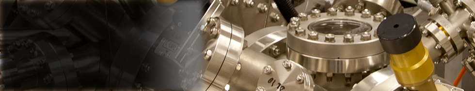
- The ultimate spatial resolution is achievable by means of the cold field emission source of electrons available in the JEOL JSM 6700F scanning electron microscope with verified resolution of 1 nm at the electron energy of 15 keV. The microscope is equipped with the energy dispersive analyser of X-rays Oxford INCA Energy 350 for chemical microanalysis.
- Methodology for the very low energy scanning electron microscopy has been developed at ISI and the corresponding attachments are installed on several microscopes in LEM. The method enables the preservation of image resolution down to arbitrarily low energy, which is not possible in commercially available instruments. At very low energies plenty of novel contrast mechanisms reveal electronic and crystallinic structures of the sample.
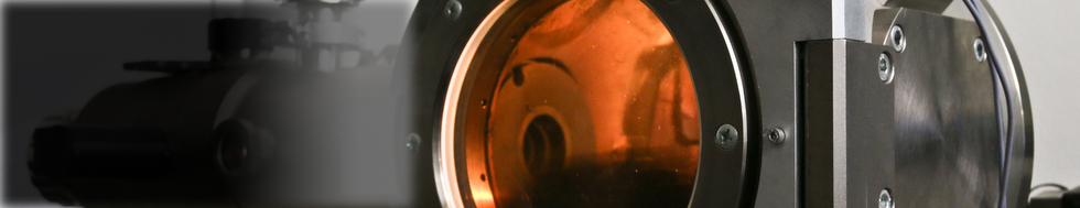
- Environmental scanning electron microscopy (ESEM) enables the study of samples of living substances and materials under elevated pressure of surrounding gas up to 3000 Pa. In this environment charging of nonconductive samples is avoided and even wet matter can be protected against drying . The ESEM instruments also work under the standard high vacuum conditions.
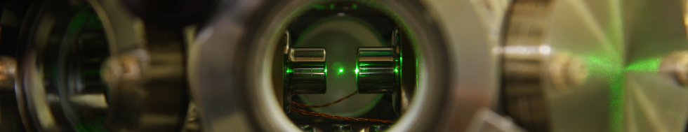
- Electron microscopy under ultrahigh vacuum conditions of 10–8 Pa enables one to study atomically clean surfaces that can be cleaned in-situ with ion beam bombardment. The device offers excellent conditions for the low energy microscopy.
- LEM has available standard preparation techniques such as sputtering and evaporation of surface coatings, ion beam thinning, exact cutting. Complementary light optical imaging is possible with the confocal microscope Olympus LEXT 3100.
Technologie:
Ing. Ilona Müllerová, DrSc.
doc. Ing. et Ing. Vilém Neděla, Ph.D., DSc.
Ing. Ilona Müllerová, DrSc.
Ing. Filip Mika, Ph.D.
Ing. Ilona Müllerová, DrSc.
Ing. Vladislav Krzyžánek, Ph.D.
Ing. Vladislav Krzyžánek, Ph.D.
